Image result for immune tolerance


----------------------------------------------------------------------------------------------------------------------------------------------------------

*** SPECIAL NOTE FROM DR. WICHMAN ***

The following excellent article was reproduced from Human Vaccines & Immunotherapeutics at https://www.ncbi.nlm.nih.gov/pmc/articles/PMC3899137/:


Hum Vaccin Immunother. 2013 May 1; 9(5): 1032–1038.
Published online 2013 Jan 28. doi:  10.4161/hv.23685
PMCID: PMC3899137

Tolerogenic vaccines for Multiple Sclerosis


Immunogenic and Tolerogenic Vaccines: A Primer

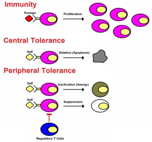
The classic immunogenic vaccines represent one of the most successful and cost-effective interventions in the history of medicine. The field of vaccination is now rapidly expanding to encompass a new class of vaccines (i.e., the tolerogenic vaccines) that induce immunological tolerance to alleviate the inflammatory autoimmune diseases as well as other inflammatory diseases of metabolism, neurodegeneration, allergic hypersensitivity, and transplantation rejection. Immunogenic and tolerogenic vaccines differ in many fundamentals. First, immunogenic vaccines have had unparalleled clinical success, including the elimination of smallpox and the near-eradication of polio and are credited with saving untold millions of lives. Conversely, tolerogenic vaccines are largely experimental and are currently pursued in pre-clinical settings or in early-phase clinical trials and are studied based on their future potential rather than their clinical actuality. Second, classic immunogenic vaccination is based on the activation, expansion, and differentiation of memory T cell clones specific for particular infectious antigens that then provide rapid immunity to any re-exposure of that infectious pathogen. Likewise, tolerogenic vaccines are contingent upon the generation of long-lasting memory, but this memory is due to the activation, expansion, and differentiation of antigen-specific regulatory T cells that mediate antigen-specific inhibitory activity by mechanisms of active, dominant tolerance. Tolerogenic vaccines also elicit ‘passive’ tolerance marked by immunological anergy and the functional and/ or physical elimination of pathogenic T cell clones and the consequent creation of a “hole in the repertoire” marked by the passive lack of T cell reactivity to a given self antigen. Third, immunogenic vaccines usually require strong immunological adjuvants to prime the initial immunization via stimulatory interactions with innate immune receptor systems that recognize “pattern-associated molecular patterns” or PAMPs that are shared among large classes of infectious pathogens. Activation of receptor systems such as the toll-like receptors (TLR) or NOD-like receptors (NLR) on mononuclear phagocytes triggers immunogenic presentation of the associated antigens on MHC class II molecules together with strong co-stimulation to elicit activation and differentiation of naïve T cells as the founding event in the adaptive immune response. In contrast, tolerogenic vaccines are designed to have minimal adjuvant activity so that the vaccine antigens are recognized in non-activated, non-inflammatory environments that favor self-tolerance. Fourth, immunogenic vaccines in current practice are largely based on the generation of humoral immunity with an emphasis on disease prevention rather than therapy of pre-established disease. In contrast, tolerogenic vaccines must counter ongoing pathogenic T cell-mediated memory, and the emphasis is on therapy of established chronic disease rather than prevention of future disease.

Tolerogenic Vaccination for Multiple Sclerosis (MS): The Targets, the Challenges, the Strategies and the Future

The use of experimental autoimmune encephalomyelitis (EAE) as the prototypic model of MS has fundamentally shaped the core expectations and predictions of MS,1-9 including the presumption that MS is an inflammatory autoimmune disease mediated by pathogenic CD4+ T-helper cells specific for myelin proteins of the central nervous system (CNS) such as myelin basic protein (MBP), proteolipid protein (PLP), and myelin oligodendrocyte glycoprotein (MOG). Myelin-specific CD4+ T cells may also recruit additional effector subsets with shared anti-myelin protein reactivity such as B cells and CD8+ T cells to partake in the autoimmune destruction of the CNS target tissue.10-16 The purpose here is not to debate the merits of the EAE model but rather to discuss the essential considerations inherent to this model for developing tolerogenic vaccines for MS. Many tolerogenic vaccine platforms have been tested for development in EAE and MS.17-29 These vaccine strategies for MS require consideration on three levels, including; (a) the identity and diversity of target CNS antigens, particularly the peptide epitopes of major myelin protein antigens, (b) the inherent efficiency by which these clones recognize their target self antigens, and (c) the qualitative activity of the vaccine in the midst of a pro-inflammatory local environment.

The Vaccine Target(s) in MS

‘Molecular mimicry’ is the leading model to explain the etiology of MS.30-33 This model predicts that chronic or recurring infectious agents are the etiological instigators of MS. The prediction is that such etiological agents have overlapping and partially shared antigenic epitopes with major myelin proteins. In the context of chronic or recurrent infection, these infectious pathogens are postulated to stimulate strong T cell-mediated immunity against the foreign “mimicry” epitope(s). After clonal expansion and differentiation into memory/ effector subsets, through mechanisms of immune surveillance, these crossreactive T cell clones are postulated to find and react to the endogenous crossreactive myelin epitopes in the CNS to mediate MS (Fig. 1). These infectious agents, upon repeated re-emergence from latency, may drive relapses of MS and the chronic relapsing-remitting disease course of MS.

figure hvi-9-1032-g1
Figure 1. Hypothetical model of action for tolerogenic vaccines. Natural homeostatic mechanisms of self tolerance are noted in blue. Endogenous myelin autoantigens and other crossreactive self antigens shape development of the T cell repertoire ...

The “molecular mimicry” model gives rise to a range of possibilities in regard to the nature of the autoantigens that drive the disease process. The key crossreactivities may be chance occurrences, and only one or two crossreactive epitopes may be responsible for initiation of the MS disease process. Conversely, the molecular mimicry may be driven by directed evolutionary pressures by which the infectious agent has adapted to a specialized infectious niche. For example, the infectious agent may be evolutionarily adapted to persist in an organ such as the CNS by bearing camouflage comprised of many epitopes that resemble and are crossreactive with the local host tissue. Mechanisms of ‘epitope spreading’ may further complicate vaccine development.34-36 “Epitope spreading” refers to a mechanism by which the instigating myelin-reactive clones drive CNS inflammation and recruit new, distinct clonotypes of naïve myelin-reactive clones into the CNS, where the latter clones differentiate into memory/ effector T cells upon recognition of a distinct set of CNS antigens to cause additional neurologic damage. Thus, over time, the anti-myelin T cell repertoire may broaden and diversify, and may acquire sufficient clonotypic diversity to sustain a chronic or progressive autoimmune course independent of any recurring infectious disease trigger.

These considerations underlie key unanswered questions that will shape the future of tolerogenic vaccine research in MS. Can effective tolerogenic vaccination be accomplished by targeting a limited number of myelin epitopes or will such vaccines need to cover a wide diversity of antigenic targets? A parallel question is the degree of patient-to-patient variability of the antigenic specificities that drive MS. The potential answers to these questions cover a wide range of possibilities, including the possibility of a common MS vaccine bearing a limited number of dominant myelin epitopes that will be effective for most MS patients. On the opposite side of the spectrum, MS vaccines may need to be individualized vaccines that cover numerous and highly diverse myelin epitopes based on unique sets of reactivities for each patient. This range of possibilities addresses important issues of feasibility and experimental approach in the design and testing of experimental vaccines. Whether pauci-clonal or broadly polyclonal T cells mediate MS is a critical consideration for tolerogenic vaccine development. If MS is mediated by T cells of restricted clonotypic diversity and little patient-to-patient variability in myelin specificities, then standard tolerogenic vaccine strategies may suffice for many MS patients. However, if MS is mediated by an ever-broadening repertoire of autoreactive T cells with substantial inter-patient variation, then new vaccine strategies will be needed to match therapy with a dynamically changing landscape of T cell autoreactivities. If the latter scenario is the case, early diagnosis and intervention will be key considerations for effective tolerogenic vaccination. New technologies will be needed to identify clonally-expanded, ‘smoking-gun’ myelin-reactive clones. Because one will not want to introduce vaccines that are off-target and not relevant to a given patient, vaccines will need to have a modular design so that epitopes can be readily added or subtracted from a vaccine cocktail to tailor the therapy to the reactivities of particular patients. Alternatively, new vaccine strategies could be developed to include full-length myelin proteins, including hydrophobic transmembrane domains. Most tolerogenic vaccine strategies have focused on defined encephalitogenic epitopes that are hydrophilic for practical reasons of solubility. Vaccines that include full length myelin proteins would need to be produced by technologies that do not result in protein aggregation, because aggregated proteins would likely cause the adverse side-effect and the strongly contraindicated production of anti-myelin autoantibodies. This spectrum of possibilities will shape the future development of optimal tolerogenic vaccines. The assumption of a common dominant MS antigen can serve as the basis to test experimental strategies designed to maximize the efficacy of tolerogenic vaccines. The assumption of myriads of vaccine targets with substantial inter-patient variation would be a compelling rationale to test novel modular vaccine designs or approaches based on full-length myelin proteins.

Antigen Recognition Efficiencies

The second consideration is the efficiency by which the myelin-reactive T cells recognize the relevant crossreactive foreign and self antigens (Fig. 1). The general consideration is that the crossreactive clonotypes recognize the foreign ‘mimicry’ epitopes of the infectious agent with qualitatively superior efficiency compared with self myelin epitopes.37-39 The differential reactivity is simply a consequence of self tolerance induction during development of the immune system, which precludes clones with strong reactivity to self.40The strength of foreign antigen recognition is considered sufficient to drive activation and the differentiation of naïve T cells into memory or effector T cells. In contrast, the less efficient myelin epitopes are postulated to lack the capacity to drive differentiation of naïve T cells. Thus, the foreign mimicry epitopes may provide the critical ‘activation energy’ to initiate autoimmune responses. The relevant myelin-reactive clones, even after achieving effector status, would continue to recognize endogenous myelin epitopes with low efficiency. Once the foreign mimicry antigen caused effector cell formation, the low efficiency recognition of myelin by effector/ memory T cells is postulated to be sufficient to elicit tissue damage39,41 but may be insufficient to sustain the immune response. Indeed, the low affinity recognition of myelin epitopes may be inherently tolerogenic.

The myelin-specific regulatory T cell networks that were overridden by the strong foreign stimulus are thought to be intact and capable of restoring homeostasis once the foreign mimicry has been cleared from the tissue (Fig. 1). These regulatory networks are most likely dominant in the absence of the infectious stimulus. The important concept is that the repertoires of the myelin-reactive regulatory T cells and the myelin-reactive conventional T cells most likely share a common myelin reactivity (representing the normal mechanism of homeostasis), but the regulatory T cells most likely do not recognize the foreign mimicry antigens due to the absence of these foreign antigens during development. If regulatory T cells per chance did crossreact with the mimicry antigens, then the immune system would not respond to these foreign mimicry epitopes, and the individual would not suffer from an autoimmune disease. Because tolerogenic vaccines contain native myelin epitopes, tolerogenic vaccination would predictably reinforce regulatory T cell function and the low efficiency interactions that promote tolerance rather than immunity, even after differentiation of the conventional myelin-reactive T cells into memory and effector subsets.

As a case in point, one of the most profoundly tolerogenic vaccines studied by our laboratory is comprised of the cytokine GM-CSF fused to the major encephalitogenic epitopes from MBP, MOG, or PLP in rat and mouse models of EAE.18,42,43 The GMCSF-neuroantigen (NAg) vaccine requires covalent linkage of the cytokine and NAg domains for tolerogenic efficacy. Conversely, a substantial number of studies have shown that GM-CSF fusions with foreign antigens act as immunogenic vaccines and can be used to augment immune responses against foreign antigens.44,45 Taken together, these studies suggest that the antigenic domain aside from the GM-CSF domain may bias the inherent activity of the vaccine. Thus, the self or nonself origins of the antigenic domain may play an important role in controlling the balance between tolerance and immunity. An important functionality of the GM-CSF domain may simply involve targeting of the vaccine to dendritic cells to promote more efficient presentation of the antigenic domain.43 The ‘self’ NAg domains of GMCSF-NAg fusion proteins, by engaging developmentally established regulatory networks, would be predicted to promote tolerance. Conversely, the ‘foreign-antigen’ domains of GMCSF-antigen fusion proteins would predominantly engage a repertoire of conventional T cells rather than regulatory T cells and thereby would drive immunogenic responses. These predictions are consistent with the experimental observations that GM-CSF may amplify the intrinsic tolerogenic or immunogenic activity of the antigenic domain. The inherent tolerogenic activity of self antigens, even in the midst of an ongoing autoimmune disease, may appear paradoxical but is a key element in the feasibility and potential success of tolerogenic vaccines.

The Environment

One of the primary concerns regarding tolerogenic vaccines is the possibility that administration of a ‘tolerogenic vaccine’ to particular patients may be immunogenic rather tolerogenic and worsen the MS disease course, as may have occurred in clinical trials of “altered peptide ligands or APL”.46 Underlying this concern is the concept that tolerogenic vaccines have tolerogenic activity because these vaccines are administered without adjuvants and thus are presented to the immune system in a quiescent, non-activated environment.47-52 The idea is that antigen presentation in the absence of costimulatory molecules and without a complement of proinflammatory cytokines is critical for inhibitory vaccine activity. However, the environment of the vaccine recipient will be difficult to control in a clinical setting because at least some patients will have inapparent infections or exposure to other pro-inflammatory stimuli either locally or systemically. If a tolerogenic vaccine is administered into such a pro-inflammatory environment, the vaccine would perhaps adversely worsen the disease course. The extent to which this issue applies to a tolerogenic vaccine platform represents a critical consideration regarding the projected success of that vaccine platform.

The notion is widely-held that a vaccine is either tolerogenic or immunogenic based on whether the vaccine is administered into a quiescent or pro-inflammatory environment. However, this idea can be challenged on theoretical and experimental grounds and may differentially apply to distinct tolerogenic vaccine platforms. Autoreactive T cells circulate in normal individuals but do not cause autoimmunity in the vast majority of persons even though the host is continually subjected to infectious disease, injury, and various inflammatory insults. Regulatory networks apparently are not so simply co-opted by inflammatory environments. Indeed, regulatory networks comprised of Foxp3+CD4+CD25+ may be strengthened by antigen recognition in the presence of B7-mediated costimulation and various cytokines, particularly IL-2.53,54 Thus, one cannot conclude that the inhibitory activity of a tolerogenic vaccine would be preempted within a pro-inflammatory environment. As a case in point, a GMCSF-NAg tolerogenic vaccine was administered at the same time and at sites immediately adjacent to the encephalitogenic peptide/ Complete Freund’s Adjuvant (CFA) immunization. In this paradigm, GMCSF-NAg retained inhibitory activity and prevented EAE in a majority of immunized mice.18 Thus, the GMCSF-NAg vaccine retained inhibitory activity even in the midst of a CFA-polarized inflammatory environment. Thus, a critical test of a tolerogenic vaccine platform is the deliberate introduction of the vaccine in a proinflammatory environment, either administered after disease onset or together with adjuvants or other pro-inflammatory stimuli during encephalitogenic immunization. In this regard, GMCSF-NAg effectively inhibited disease when delivered into the same lymphatic drainage at the same time as the encephalitogenic/ CFA immunization. The GMCSF-NAg vaccine also inhibited EAE when given at disease onset, or alternatively, when given during established chronic EAE.18,42,43

The Future

A primary prediction of the EAE model of MS is that the study of the myelin-specific T cell repertoire of MS patients should reveal the key specificities that underlie MS. However, many studies have shown that MS patients had similar frequencies of myelin reactive T cells compared with healthy age- and gender-matched controls.55-61 The myelin-specific repertoire in MS nonetheless appears to show some evidence of clonal expansion and a more pronounced expression of an activation/memory phenotype. The ‘cause or consequence’ question however has yet to be answered, and no clear consensus has emerged regarding the diversity and inter-patient variability of the antigenic determinants that drive the MS disease process. The questions of which peptides cause pathogenesis in MS are particularly vexing. A clinical trial of an ‘altered peptide ligand’ of MBP83–99 was designed to inhibit MS but instead appeared to aggravate disease.46 This adverse effect provided suggestive evidence implicating this myelin peptide as encephalitogenic in humans. Other CNS proteins aside from the myelin antigens might be critical antigenic targets in MS,62-66 just as aquaporin-4 appears to be a major antigenic target in Neuromyelitis Optica, and the inward-rectifying KIR4.1 potassium channel appears to be a major target of the humoral immune response in many adult MS patients. Humoral responses to MOG are apparent in patients with Acute Disseminated Encephalomyelitis and pediatric MS. Human T cell antigen receptor chains specific for myelin peptides have been expressed in mice as transgenes together with the human MHC class II restriction alleles, and these transgenic mice show spontaneous EAE.67-69 These and other approaches provide compelling evidence that particular antigen specificities are associated with MS and related neurologic diseases. However, the true encephalitogenic drivers of MS remain unknown. As a key question for the future of the field, consideration must be given to those experimental approaches that might best reveal causal links between a given antigenic specificity and MS pathogenesis. Tolerogenic vaccines may provide the key to the solution. In this regard, a central goal of the field is to devise and test tolerogenic vaccines as interventions in MS. A major step forward would be a successful clinical trial of an efficacious tolerogenic vaccine. Such an accomplishment would verify the tolerogenic efficacy of the vaccine approach, and the identity of the vaccine NAg domain would definitively link that NAg specificity as a foundation of MS pathogenesis. Thus, clinical trials of tolerogenic vaccines may be a key experimental venue to not only treat MS but to reveal the underlying pathogenesis of MS. Thus, over the course of the next decade, the hope is that tolerogenic vaccines will begin to reveal the “nature of the beast” that is MS.


Articles from Human Vaccines & Immunotherapeutics are provided here courtesy of Taylor & Francis


*** This page is under construction. If you would like to know more about this topic, please call 770-232-7883 or contact us by This email address is being protected from spambots. You need JavaScript enabled to view it..

Image result for low dose allergens


LDA Therapy an environmental medicine therapy invented by W. A. Shrader, MD. LDA is also known as low dose allergen therapy or ultra low dose enzyme activated immunotherapy. It is an immunotherapy enhanced by a small dose of the enzyme beta glucuronidase. The enzyme activates extremely miniscule doses of various allergens and stimulates the production of T Regulatory cells. These cells “switch off” helper cells that are erroneously causing patients to be ill by misidentifying normal substances in the body to be allergens. T-cells may remain active for long periods of time in the bloodstream, so LDA will need to be administered only every 2 months at first, and then less often as time passes.

LDA is used to treat all types of allergy, sensitivity and intolerance to inhalants, foods and chemicals. It is used to treat such conditions as hay fever, asthma, all types of food allergy and many other problems listed on Dr. Shrader’s website.

LDA Hisory

LDA is patterned after the Enzyme Potentiated Desensitization (EPD) immunotherapy method. The EPD method involves desensitization with combinations of a wide variety of extremely low dose allergens (1 part in 10 million to as low as 1 part in 1 quadrillion). These allergens are given with beta-glucuronidase. The beta-glucuronidase acts as a lymphokine, a substance that potentiates the immunizing ability of the allergens. The EPD method appears to induce the production of activated T-regulator cells, which can live in circulation for many years.

The use of LDA is limited by necessity in the USA because it is available only by prescription for specific physicians’ patients and is not available as a retail product.

LDA Compared to Conventional Allergy Immunotherapy

With conventional “escalating dose” immunotherapy, the dose is started “low” (generally 1 to 10,000, and then increases over time to as high as 1 to 10, 1 to 20 or 1 to 100). Conventional allergy therapy is practiced primarily to treat IgE mediated allergic reactions. This type of immunotherapy works by causing the patient to produce blocking IgG antibody, which inhibits histamine-releasing ability (the allergy symptoms).

To produce adequate levels of blocking antibody, very high doses of allergen are given. This can be dangerous because of the risk of severe reactions. Deaths from conventional escalating dose immunotherapy are generally a result of anaphylaxis. LDA immunotherapy, however, is cell-mediated and extremely low dose. The very highest dose of LDA is at least a million times less than the standard dose for conventional immunotherapy. Life-threatening reactions to EPD or LDA have never been reported.

Conventional escalating dose immunotherapy is generally administered twice weekly for the first four to six months of treatment. Once the very high maintenance dose is reached, the treatment interval may be extended to once every two weeks or monthly, but rarely less often without return of symptoms. Conventional escalating dose immunotherapy cannot usually be stopped without the return of symptoms within 3 to 12 months of cessation.

LDA is administered every other month for the first year. After the initial 12-month period, the treatment interval may generally be extended to three months or longer. Most adults with significant problems require 16 to18 treatments at two-month intervals, at which time treatment often may be discontinued. Of the approximately 50% of patients who are unable to discontinue LDA after 16-18 treatments without return of some symptoms, the majority will continue treatment longer at intervals of 6 months to a year. While LDA is effective with children, our office does not treat children. Dr. Shrader’s office does treat children.

LDA Treatment

A majority of LDA patients (over 60%) note a significant positive response to LDA with their first treatment. Most other patients respond positively by the third treatment. A small percentage of patients do not have strongly positive results until they have had 6 treatments. The overall failure rate (no improvement) is about 9%.

Available LDA mixtures include 1) inhalants (inhaled pollens, animal danders, dust and mites, insects, fungi, and yeast, including candida species, and molds), 2) foods and food additives, 3) chemicals (containing most common chemicals and scents, formaldehyde and detergents, except for pesticides and herbicides), 4) woods (a mixture of over 90 common and exotic woods), used for the treatment of contact skin sensitivity in woodworkers, and 5) recently added Lyme.

Other specific LDA mixtures are available to treat several specific autoimmune diseases, such as rheumatoid arthritis, ulcerative colitis, ankylosing spondylitis, scleroderma and others. These mixtures work by a mechanism called molecular mimicry.

There are some disadvantages to doing LDA. For example, the day before, the day of, and the day after LDA treatment most patients must adhere to a very restricted diet. There are also medications (such as antihistamines and aspirin) that may significantly reduce or destroy the effectiveness of LDA if taken in the three weeks after treatment. An LDA booklet is provided to patients to ease compliance.

LDA is dramatically effective for the treatment of eczema. Likewise, immediate food allergy, which can cause life-threatening anaphylaxis, has no effective treatment except for emergency drug treatment and avoidance of the offending food. LDA practitioners report that LDA appears to work well for this condition.

Overall, LDA immunotherapy is considered an extremely beneficial treatment by thousands of patients across the US and Canada who rely on it for treatment of a myriad of problems.

Note: LDA is not approved by the Food and Drug Administration.

Source: http://www.drshrader.com/

See also: www.nwhealthcare.net/index.php?id=69

Healing from the “Sunshine Vitamin” Vitamin D Promotes Strong Bones, Strong Immune System

Vitamin D is important for strong bones, teeth and immunity system. Many decades of studies have proven that Vitamin D is vital for bone and muscle growth and the regulation of other minerals in the body. Our own skin produces Vitamin D when exposed to the Sun.However, with fewer people going outside or using sunscreen and eating Vitamin D rich foods like fatty fishes (salmon, herring, tuna, sardines) and eggs, many people are feeling the effects of Vitamin D deficiency. Many health authorities suggest at least 400 IU’s per day, but others believe that is woefully inadequate and suggest up to 4,000 IU’s per day.


Why Vitamin D is Important

Vitamin D promotes bone growth by allowing calcium to travel through cell membranes and reach the bones. It also controls calcium in the bones and helps the body absorb magnesium, iron and zinc. If your body doesn’t find enough Vitamin D, it will take it from the bones and this can contribute to osteoporosis and other bone-weakening diseases.Also, researchers have discovered that proper Vitamin D levels can boost the immune system and muscle strength.Vitamin D triggers the body’s immune cells to produce antibodies, which promotes a strong immune system.Some studies have suggested that Vitamin D surpasses Vitamin E as an antidioxidant.Vitamin D3, also known a cholecalciferol, is its most effective form and is crucial to the proper control of calcium in the body.

Lack of Vitamin D can contribute to the following conditions.

  • Osteoporosis and Osteomalacia
  • Greater risk of diabetes
  • Compromised immune system
  • Infertility
  • Fatigue
  • Higher risk of breast, prostate, skin and colon cancer
  • Risk of stress
  • Risks of Multiple Sclerosis
  • Depression

Our Solution:

Vitamin D3 Intramuscular Therapy is a simple injection of this important supplement. This procedure is done in our office by our qualified staff of RN’s and Therapists will not take up much of your time. Also the benefits can be felt within hours of the treatment. You will have more energy and a vastly improved immunity system.

Vitamin D3 IM Therapy Benefits:

  • Stronger bones and muscles
  • Improved overall health
  • Improved immunity system
  • Decreased risk of osteoporosis
  • Improved mood

In order to qualify for the Vitamin D3 IM Therapy:

  1. Calcium, Phosphorus blood levels must be taken to rule out Hypercalcemia, parathyroid issues.
  2. Vitamin D blood level must be taken to rule out Vitamin D toxicity.
  3. PTH may also be taken in some cases to rule out parathyroid issues.
  4. Kidney and liver function needs to be checked to rule out failures respectively.

Resources:

Vitamin D and calcium supplementation reduces cancer risk: results of a randomized trial – American Journal of Clinical Nutrition

The Miracle of Vitamin D – The Weston A. Price Foundation

Vitamin D Therapy Halts Cancer Growth and Supports Immune Function – American College for Advancement in Medicine

Vitamin D shown to cut cancer risk in women – MSNBC.com

Unraveling the Enigma of Vitamin D – The National Academy of Sciences

Vitamin D the Natural Way to Prevent and Treat Flu Including Swine Flu! – Alliance for Natural Health’, ‘

Image result for estriol


----------------------------------------------------------------------------------------------------------------------------------------------------------

*** SPECIAL NOTE FROM DR. WICHMAN ***

The following excellent article was reproduced from the International Journal of Biomedical Science at https://www.ncbi.nlm.nih.gov/pmc/articles/PMC3614637/:


Int J Biomed Sci. 2006 Dec; 2(4): 305–307.
 
PMCID: PMC3614637

Estriol and Progesterone: A New Role for Sex Hormones


INTRODUCTION

It has been increasingly apparent that effects of sex hormones extend far beyond their predominant role in sexual differentiation and reproduction. Sex hormones appear to be involved in regulating the immune response, which is evident from a striking difference between a male and female immune response, and the alteration of female immune response during pregnancy, lactation and different phases of menstrual cycle (1). There is a distinct female preponderance of most autoimmune diseases, as well as a gender dimorphism related to a disease progression of autoimmune diseases. Moreover, numerous in vivo studies reported that course and severity of certain autoimmune diseases are modulated not only by castration or sex steroid administration, but also by pregnancy and lactation (2).

Immune response is regulated by components of innate immunity, as well as by components of acquired immunity, such as T helper (Th) cells subdivided to Th1 and Th2 subsets. A third Th cell subset, a regulatory T cell (Treg), has been recently identified. Th1 cells primarily secrete pro-inflammatory cytokines as IFN (interferon)-γ, IL (interleukin)-2 and TNF (tumor necrosis factor)-α, which promote cellular immunity, whereas Th2 cells secrete a different set of anti-inflammatory cytokines IL-4, IL-5, IL-6 and IL-13, which promote humoral immunity. Treg cells produce cytokines like IL-4, IL-10 and TGF (transforming growth factor)-β, and have suppressive properties for both Th1 and Th2 cells. A Th1 and Th2 responses are mutually inhibitory, and to some extent opposing, whereas Treg prevent the derangement of the Th1/Th2 balance in either direction (34).

ESTRIOL AND PROGESTERONE: IMMUNITY AND NEUROPROTECTION

Copious data support the concept of an altered Th1/Th2 balance during pregnancy, suggesting that pregnancy can be regarded as a Th2-type phenomenon. A dominance of Th-2 type cells prevents a rejection of antigenically foreign fetus by a cell-mediated immune attack (5). Pregnancy-associated Th2 shift has been proposed as a mechanism underlying the improvement of Th1-mediated autoimmune diseases (as rheumatoid arthritis, multiple sclerosis (MS), autoimmune thyoriditis, uveitis, and psoriatic arthritis), or a deterioration of Th2-mediated autoimmune diseases (as systemic lupus erythematosus).

A remarkable feature of pregnancy is a successful adaptation of woman to enormous endocrine changes as increased production of estrogens, progesterone (PRG), corticosteroids, and numerous protein hormones (some are placental specific and unique to pregnancy) (6). During pregnancy PRG serum level increases by a factor of 4, while estrogen estriol (E3) serum concentration increases by a factor of 20. Normally, E3 is not measurable in serum. E3 is produced principally from 16α-hydroxydehydroepia ndrosterone sulfate in the fetal plasma. Its level rises during pregnancy, peaking in the third trimester, and drops postpartum. In the urine of nonpregnant women the ratio of E3 to E1 (estron) plus E2 (estradiol) is approximately 1:1, whereas in the urine of pregnant women the ratio is 10:1 or more. In pregnant women at or near term there is a daily production of about 300 μmol (80 mg) of E3 and 1 mmol (300 mg) of PRG (6).

Estrogens and PRG induce a substantial Th2 shift, most likely at concentrations associated with pregnancy.

The estrogens are known to exert opposing, bimodal, dose-specific effect to immune response. Low levels facilitate a cell-mediated pro-inflammatory immune response, whereas their relatively high levels, such as those achieved during pregnancy, promote an anti-inflammatory Th2 response (7). Studies in the last decade showed that estrogens in high concentrations have a beneficial effect on the experimental autoimmune encephalomyelitis (EAE), a mouse model of chronic relapsing-remitting form of human MS. In a recent EAE study, a long-term treatment of castrated female mice with E3, given at doses corresponding to those seen during late stage of pregnancy, delayed the disease onset even for a longer time than treatment with E2 (8). Autoantigen myelin basic protein (MBP)-specific T-lymphocyte responses from E3-treated EAE mice are characterized by significantly increased production of cytokine IL-10 (9). Moreover, it has been reported that estriol’s ameliorative effect is not gender-specific. E3 treatment decreased the EAE disease severity, as well as pro-inflammatory cytokine production in both females and males (10). Furthermore, in a pilot trial, oral E3 treatment of women with relapsing-remitting MS (RRMS) caused significant decreases in the number of gadolinium-enhancing lesions on brain magnetic resonance imaging (MRI), and significant increases of IL-5 and IL-10 levels. These changes in cytokines correlated with reductions of enhancing lesions on brain MRI (1112). In women with secondary progressive MS (SPMS) a lack of treatment effect is believed to support a concept of different immune dysregulation.

Estrogens can display a wide range of neuroprotective activities, including stabilization of neurotransmission, inhibition of apoptosis, and direct antioxidant activities which may be important for defense against oxidative stress (13). Similarly, physiological concentrations of PRG and estrogens, consistent with late pregnancy, are shown to inhibit activated microglia, resident central nervous system (CNS) cells associated with a wide variety of neuroimmunological diseases, including MS. In addition, PRG and estrogens inhibited microglial production of NO (nitric oxide) and TNFα, molecules that can be toxic to myelin-producing oligodendrocytes and neurons (1415).

PRG mediates promotion of Th2 response, inducing conversion of Th0 cells into Th2 cells. At concentrations comparable to those present at the maternal-fetal interface PRG induces a development of Ag-specific CD4+ T cell lines that show enhanced ability to produce IL-4 and IL-5 (16). Also, PRG directly suppresses T cell differentiation into Th1 cells, and enhances IL-10-producing Th2 cells (17). On the other hand, PRG has been shown to provide substantial neuroprotective effect in male and female animals after brain or spinal cord injury. As an acute post-injury treatment PRG reduces cerebral edema, neuronal loss, and inflammation process (18-20). In a recent pilot clinical trial using PRG to treat traumatic brain injury (TBI) patients, stable PRG concentrations were rapidly achieved following continuous intravenous infusion, whereas alterations in PRG pharmacokinetics were not gender specific. Also, PRG showed possible signs of benefit with no appreciable effects on heart rate, coagulopathy or infection rate, important considerations in seriously injured patients (2021).

CONCLUSION

Taken together, the above studies imply that the effects of sex hormones on the immune and nervous system are clearly apparent, although imperfectly understood (Table (Table1).1). The transition from animal and cellular models to human therapies is fraught with difficulty, especially concerning some large clinical trials suggesting that some hormone therapies may increase the risk of several diseases, including stroke. The results of these studies may be due to the doses, specific hormone used, and sub-population of patients. However, a several clinical trials on the effects of sex hormones to MS or TBI have been performed. Their results suggest that hormone therapy may represent a new therapeutic tool to combat certain diseases. Estriol, the safest of three estrogens, is a candidate for treatment women with RRMS, based on a recent phase I clinical trial. Since estriol’s ameliorative effect is not gender-specific, the possibility of estriol treatment in men exists. In a pilot trial, PRG has already been used in both sexes of patients with TBI.

Table 1
A summary of experimental and clinical studies on the effects of pregnancy-related sex hormones

Thus, further randomized clinical trials are necessary. It is important to minimize the risk of treatment, and design therapies optimized for anti-inflammatory and neuroprotective efficacy.


Articles from International Journal of Biomedical Science : IJBS are provided here courtesy of Master Publishing Group

Image result for leaky gut dysbiosis


Stress, bowel disorders and a poor diet can damage the cellular lining of the intestines, allowing toxins, bacteria, waste and food particles to leak through. Leaky gut syndrome, also known as intestinal permeability, may lead to bowel disorders, poor nutrition, Crohn’s disease, and inflammation.

.The intestinal walls are lined with villi, which absorb nutrients through Tight Junctions. However, as the villi become more permeable, large proteins slip into the bloodstream and triggers an immune response, which can then cause food allergies and further inflammation. Tests exist that detect the symptoms of leaky gut.

The Lactulose Mannitol Test (LT) or Polyethelyne Glycol (PEG) test administers two sugars a lactulose solution and mannitol into the body. If test shows a higher than normal lactulose and mannitol in the body, this means the body is not absorbing them, which is an indicator of leaky gut. The doctor can follow up with a treatment regimen to reduce inflammation.

Gut dysbiosis is an imbalance in the microecology of the digestive tract that can cause abdominal pain, bloating and distention, flatulence (gas) and diarrhea. When the microbial balance of the gut is disturbed, opportunistic, (“bad”) bacteria can overgrow the gut’s good bacteria. At the Advanced Rejuvenation Institute, we use the Hydrogen Breath Test in the diagnosis of three primary conditions that can cause gut dysbiosis:

  • Improper digestion of sugars: The most common sugar that is poorly digested is lactose, the sugar in milk (lactose intolerance). Testing also may be used to diagnose problems with the digestion of sucrose, fructose and other sugars.
  • bacterial overgrowth of the small bowel, a condition in which larger-than-normal numbers of colonic bacteria are present in the small intestine.
  • if individuals have abnormally rapid passage of food through the small intestine, there may not be enough time for the small intestine to digest and absorb sugars and carbohydrates. This results in the entry of larger amounts of sugar and carbohydrate into the colon where the bacteria can digest and convert them to gas.

Bacterial overgrowth can inhibit nutrient absorption and lead to the following serious health problems:

  • Intestinal permeability (leaky gut)
  • Carbohydrate intolerance
  • Malabsorption
  • Malnutrition and weight loss
  • Anemia
  • Fibromyalgia
  • Chronic Fatigue Syndrome
  • Osteoporosis

The Hydrogen Breath Test measures levels of hydrogen in the breath to diagnose several conditions that cause gastrointestinal symptoms. In humans, only anaerobic bacteria in the colon can produce hydrogen. These bacteria produce hydrogen when they are exposed to unabsorbed sugars and carbohydrates (not proteins or fats). Large amounts of hydrogen may be produced when there is a problem with the digestion or absorption of food in the small intestine that causes unabsorbed food to reach the colon. Large amounts of hydrogen also may be produced when the colon bacteria backflow into the small intestine, a condition known as bacterial overgrowth of the small bowel. When the backflow bacteria are exposed to unabsorbed food that has not had a chance to completely traverse the small intestine to be fully digested and absorbed, some of the hydrogen produced by the bacteria, whether in the small intestine or the colon, is absorbed into the blood flowing through the wall of the small intestine and colon. The hydrogen-containing blood travels to the lungs where the hydrogen is released and exhaled in the breath where it can be measured.

Research indicates that a large number of Americans suffering from abdominal pain, bloating, diarrhea, and other guts issues, were found to have bacteria that are normally found in the large intestine backed up into the small intestine. There appears to be a strong correlation between this situation and Irritable Bowel Syndrome. Once diagnosed, the condition can be treated, patients with this condition have experienced welcome relief from their symptoms. Similarly, accurate diagnosis of one or more of the sugar intolerances can lead to development of a treatment plan that can greatly diminish painful digestive reactions to lactose, fructose, or sucrose.

The test is simple, non-invasive, patient-friendly and is performed after a short period of fasting (typically 8-12 hours).

Sources and Research:

Tight junctions, leaky intestines, and pediatric diseases

dyspepsia-symptoms

The Importance of Proper pH Balance of the Gastrointestinal System

Do you:

  • have indigestion, stomach pain or acid reflux?

  • wonder if you absorb nutrients efficiently?

  • have food allergies?

THE PROBLEM

Have you been told that you have “heart burn”, “indigestion” or “acid reflux” and that you’ll need to be on medication for the rest of your life to control your stomach pain? Does this seem right to you? Why not focus on fixing the problem rather than masking its symptoms with drugs for the rest of your life?

How did this happen?

Most people in our Western society and Western Medical Doctors are misguided about the role stomach acid plays in health. Most people:

  • Think they have too much stomach acid, when in fact they have too little.
  • Treat the “acid reflux” or “heart burn” symptom by suppressing stomach acid production, which leads to long-term health problems.
  • Eventually lose the capacity to produce sufficient stomach acid as a result of not eating enzyme-rich raw plant foods and other dietary mistakes and continual reliance on antacids that suppress the body’s ability to produce stomach acid.

You’ve spent most of your life eating cooked, processed foods that have had all of their naturally-occurring digestive enzymes destroyed. The lack of these natural digestive enzymes in your food forces your body to overproduce stomach acid to compensate for the lack of digestion that would have occurred with the enzymes. Eventually, the long-term over-production of stomach acid can cause the stomach to loose its ability to produce stomach acid.

Over the years, your body’s capacity to produce stomach acid begins to diminish. Diminished stomach acid causes the loss in your body’s ability to sufficiently process food in the stomach. The health consequences of inadequately digested food can be profound.

Low production of stomach acid has become very common. A person over age 40 who eats a Standard American Diet has a high probability of having low stomach acid. Note that symptoms of low stomach acidity are often the same symptoms associated with too much stomach acid: heartburn, bloating, belching, and flatulence, Indigestion, diarrhea, or constipation. Many people who think they are suffering from too much stomach acid are actually suffering from the exact opposite condition. The use of antacids and purple pills then become exactly the wrong treatment to use since they exacerbate the underlying condition while temporarily masking the symptoms.

-heartburn_FS

Consequences can include:

  • Poor digestion: Insufficient stomach acid means food cannot be adequately broken down. To make things worse, insufficient stomach acid also means that there is insufficient acidity to optimize the digestive enzyme pepsin, which requires a pH of around 2.0. Pepsin breaks down meat. Long-term insufficient stomach acid results in continual partial digestion of food, gas, bloating, belching, diarrhea/constipation, autoimmune disorders, skin eruptions, and a myriad of intestinal disorders .
  • It is estimated that 80% of people with food allergies suffer from some degree of low acid production in the stomach.
  • Many vitamins and minerals require proper stomach acid pH in order to be properly absorbed. These include: Vitamin B12, calcium, iron, and folic acid.
  • With low stomach acidity and the presence of undigested food, harmful bacteria and other pathogens can more readily colonize the stomach and further interfere with digestion.


What to do

This painful situation can be turned around with a little bit of diagnostic work at the Advanced Rejuvenation Institute, some reparative therapy, and some education combined with dietary changes. Here’s the scenario:

  1. Our office will conduct a Hiedelberg pH Gastrogram test on your stomach to determine its pH.
  2. If your stomach pH is low, you will be prescribed a treatment plan to optimize the stomach pH,
  3. By following the treatment plan, you’ll be able to experience the disappearance of your symptoms and will not have to rely on band aide medications.

01


How does the Heidelberg pH Capsule System work?

The Heidelberg pH Capsule is a non-radioactive, miniature transmitter that is about the size of a regular vitamin pill and is designed to be swallowed. After you swallow the capsule, the Heidelberg pH Capsule continuously transmits the pH values from the gastrointestinal tract as the capsule passes through your digestive system. The frequencies transmitted, by the capsule are picked up by the Medallion Pendant Transceiver (worn by the patient). This converts the Transmitted data to DIGITAL SIGNALS which are further transmitted to the Central Computer Interface, which is connected to the Computer and displayed on the Computer Screen. On completion of your test, the Technician will make a Print-out of your test information for Dr. Wichman to evaluate.

Our test is far better than tests given in the hospital to obtain the same information. There is a hospital procedure whereby a naso-gastric tube is introduced into the nose. The patient must cooperate by swallowing to allow the operator to penetrate the tube into the stomach. This is a drastic and unpleasant procedure. A suction device aspirates (sucks out) some of the stomach juices and the operator determines the pH with a lab procedure. Unfortunately, this drastic hospital procedure is not carried through to the small intestine where food conversions and absorptive processes take place. Fortunately, the Heidelberg pH Capsule does the complete job. The Heidelberg pH Capsule eliminates the need for the Stomach Tube. It allows us the opportunity to determine your vital pH data simply and painlessly in our office.

NO HOSPITAL ADMISSION…NO SPECIAL PROCEDURES ROOM…NO STOMACH TUBE…NO DISCOMFORT!

pH plays an all-important role in how we handle and process foods to the nourishment of our bodies. In addition, medications, which depend upon pH for release and absorption, have optimal effects under normal pH conditions. Virtually everything that we eat is properly converted and absorbed in the small intestine. Due to abnormal pH profiles in many patients, sustained-release medications may tend to dump or release medication all at once or may release very little. A normal, or near normal, pH alimentary canal profile is very beneficial for food processing and medication delivery. The Heidelberg pH Capsule is used routinely by many of the major Pharmaceutical Manufactures, in Europe, Japan, Canada and the United States, as a design tool in the development and formulation of pH-dependent drug releases.

MORE ABOUT THE HEIDELBERG pH CAPSULE SYSTEM

The patient wears a Medallion 2000 Pendant Transceiver around his/her neck via a Camera strap. The pendant picks up the telemetric pH data from the patient’s abdomen. It converts the information to digital data, and then, this information is transmitted to a Computer Interface Module (HEIDELBERG MEDALLION 2000) for signal processing. The Computer Interface transfers this Data to our office computer. The information that is received on the graph (or Gastrogram) is compared with your medical history. This graph gives our doctor more complete and essential data to evaluate in your diagnosis. Heidelberg pH Capsules are used extensively throughout the world in Pharmaceutical research, Pharmaceutical design, Preventive and Nutritional Medical Practices, and by dozens of major Pharmaceutical Manufacturers, world-wide. There have been over 143 published clinical studies in the Archives with over 73 pertinent studies published since 1977.The Heidelberg pH Diagnostic System is a State-of-the-Art Diagnostic Tool for Measuring the pH Levels in the Digestive Tract.

Hyperbaric Oxygen Therapy


Hyperbaric Oxygen Therapy (HBOT) helps all kinds of people. It’s used to help patients suffering from sports injuries, Chronic Fatigue Syndrome, infections, arthritis and a huge range of other medical conditions. As we all know, oxygen is vital for life, and life cannot exist without it. It follows that oxygen is essential for effective healing and recovery.

read more

Book Your Appointment NOW

Call (770) 232-7883


Click to E-Mail Us Now!


Our Address:

4488 N. Shallowford Road, Suite 201

Dunwoody, Georgia 30338


We Serve the Following Areas:

Atlanta, Georgia, Acworth, Alpharetta, Berkeley Lake, Braselton, Brookhaven, Buckhead, Buford, Canton, Chamblee, Conyers, Cumming, Dacula, Decatur, Doraville, Duluth, Dunwoody, Georgia, Flowery Branch, Gainesville, Grayson, Hoschton, Johns Creek, Kennesaw, Lawrenceville, Lilburn, Lithonia, Loganville, Marietta, Milton, Norcross, Roswell, Sandy Springs, Smyrna, Snellville, Stone Mountain, Sugar Hill, Suwanee, Tucker, Vinings, Woodstock, Georgia